The Tissues is part of the Anatomy and Physiology section which provides High Yield information suitable for the MCAT, an entrance exam for medical schools in the United States.
Four major tissue types
- Epithelial Tissue
- Connective Tissue
- Muscle Tissue
- Nervous Tissue
Epithelial (epithelium)
- 1. covers and lines body parts (sheets of cells)
- 2. glandular epithelium
- a. two major types
- 1) endocrine glands secrete hormones to blood (no ducts)
- 2) exocrine secrete products into ducts that open to skin or lumen of organ
- b. structural classification of exocrine glands
- 1) multicellular – form a distinctive structure or organ (e.g., sweat, salivary)
- 2) unicellular – have no ducts but still considered exocrine (e.g., goblet cells)
- c. functional classification of exocrine glands
- 1) holocrine – cell accumulates product, cell dies, bursts open and substance secreted (e.g., sebaceous)
- 2) merocrine – secrete by exocytosis (most glands)
- a. two major types
- 3. epithelial sheets – special characteristics
- a. continuous sheets of closely packed, tightly joined cells
- b. have apical (free) and basal surface
- c. attached to 2-layered basement membrane
- 1) basal lamina – proteins and polysaccharides secreted by epithelial cells
- 2) reticular lamina – protein fibers and glycoproteins secreted by underlying connective tissue
- d. avascular – exchanges occur by diffusion from blood supply of underlying connective tissue
- e. have nerve supply
- f. high capacity for regeneration (lots of mitosis)
- g. basic functions – protection, secretion, absorption
Connective tissue
- 1. special characteristics
- a. made up of living cells plus non-living extracellular matrix
- 1) -blasts are immature cells that secrete matrix (e.g., fibroblasts, chondroblasts,
osteoblasts) - 2) -cytes are mature cells that help maintain matrix (e.g., chondrocytes, osteocytes)
- 3) other cell types include macrophages, plasma cells (secrete antibodies), mast cells (store chemicals that help fight invaders)
- 4) matrix consists of protein fibers embedded in ground substance (polysaccharides and proteins); supports cells structurally and functionally
- 5) fibers include collagen (strong, flexible), elastin (strong, very stretchy), reticular fibers
(collagen with coating of glycoprotein, forms branching networks that support tissues and organs)
- 1) -blasts are immature cells that secrete matrix (e.g., fibroblasts, chondroblasts,
- b. has nerve supply, except cartilage
- c. most highly vascular, except cartilage which is avascular, and tendons/ligaments which have a low supply
- d. functions – support, protection, binding
- a. made up of living cells plus non-living extracellular matrix
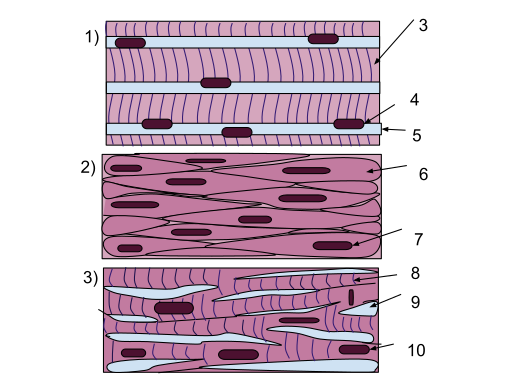
Muscle tissue
- 1. Generates force, movement, generates heat
- 2. Three types – skeletal, smooth, cardiac
- 1) Skeletal muscle cells:
- are long tubular cells with striations (3) and multiple nuclei (4).
- The nuclei are embedded in the cell membrane (5) so that they are just inside the cell.
- This type of tissue occurs in the muscles that are attached to the skeleton.
- Skeletal muscles function in voluntary movements of the body.
- 2) Smooth muscle cells:
- are spindle shaped (6), and each cell has a single nucleus (7).
- Unlike skeletal muscle, there are no striations.
- Smooth muscle acts involuntarily and functions in the movement of substances in the lumens.
- They are primarily found in blood vessel walls and walls along the digestive tract.
- 3) Cardiac muscle cells:
- branch off from each other, rather than remaining along each other like the cells in the skeletal and smooth muscle tissues.
- Because of this, there are junctions between adjacent cells (9).
- The cells have striations (8), and each cell has a single nucleus (10).
- This type of tissue occurs in the wall of the heart and its primary function is for pumping blood.
- This is an involuntary action.
Nervous tissue
- 1. initiates and transmits electrical signals
- 2. neurons and neuroglia (support cells)
Cell Junctions

LadyofHats / Public domain
- – Tight junctions
- 1. adjacent plasma membranes are fused
- 2. forms barrier that prevents leakage
- 3. common in epithelial sheets
- – Gap junctions
- 1. cells linked by protein tunnels called connexons
- 2. allow small molecules to pass between cells
- 3. important in conducting electrical signals (e.g., cardiac muscle)
- – Desmosomes
- 1. scattered over the membrane surface
- 2. found all over the body, but more common in tissues that experience stretching (e.g., skin, digestive tract)
Membranes
- – Most are epithelial membranes
- 1. epithelium with underlying CT
- – Cutaneous membrane covers body surface (skin)
- – Mucous membranes (mucosa)
- 1. line body cavities open to the exterior
- a. in respiratory, digestive, reproductive and urinary systems
- 2. form a barrier to invaders, important in body defense
- 3. tight junctions prevent leakage
- 4. secretes mucus, which moistens, lubricates, traps dust and invaders
- 5. underlying CT layer called lamina propria
- 6. formed from different kinds of epithelium
- 1. line body cavities open to the exterior
- – Serous membranes (serosa)
- 1. lines body cavities not open to exterior, covers organs
- 2. simple squamous epithelium with areolar CT
- 3. two layers
- a. parietal – attached to cavity wall
- b. visceral – covers organs
- c. between layers is serous fluid secreted by the epithelial cells
- 4. includes pleurae, pericardium, peritoneum
- – Synovial membranes
- 1. line cavities of synovial joints
- 2. no epithelium
- 3. areolar CT with elastic fibers and fat
- 4. secretes synovial fluid to lubricate joint


