Thorax

OpenStax College / CC BY
Neck Region
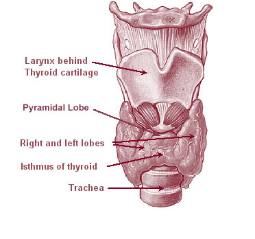
Thyroid
Esophagus
| Location | Arterial Supply | Venous Supply |
| superior esophagus | inferior thyroid artery | |
| middle esophagus | intercostals | intercostals drain into azygous |
| lower esophagus | gastric, bronchial, and splenic arteries | |
| proximal esophagus | subclavian and brachiocephalic veins | |
| distal esophagus | IVC, hepatic portal veins, gastric veins |
Abdomen
Abdominal Arteries and Veins

OpenStax College / CC BY
| Superficial arteries of the abdomen | Branches of femoral artery supply tissue above external oblique. |
| Deep arteries of the abdomen | The inferior epigastric is supplied by the external iliac. The superior epigastric is supplied by the internal thoracic |
| Venous supply of abdomen above the umbilicus | Internal mammary vein |
| Venous supply of abdomen below the umbilicus | Drain into the external iliac vein |
Arcuate Line
Anterior abdominal Layers
1. Skin
2. Subcutaneous fat
3.Superficial fascia
a. Camper’s fascia -> superficial fatty layer
b. Scarpa’s fascia -> deep membranous layer
4. Deep fascia -> Rectus sheath
5. Rectus abdominis, pyramidalis
6. External oblique muscle
7. Internal oblique muscle
8. Transversus abdominus muscle
9. Transversalis fascia
10. Preperitoneal fat
11. Parietal peritoneum
3 anterior abdominal wall folds
Stomach

Jiju Kurian Punnoose / CC BY-SA
Liver
| Arterial Supply | Venous Supply |
| Hepatic artery (30%) and portal vein (70%). 1.5L/ min | Right hepatic vein drains to IVC and middle and left hepatic veins join outside the liver. |
| Ligamentum venosum | a fibrous remnant of the ductus venosus Lies in the fissure on the inferior surface of the liver, forming the left boundary of the caudate lobe of the liver. |
| Falciform ligament | sickle-shaped peritoneal fold connecting the liver to the diaphragm and the anterior abdominal wall. |
| Ligamentum teres hepatis | contains remnant of the left umbilical vein which carries oxygenated blood from the placenta during fetal life. in the free margin of the falciform ligament and ascends from the umbilicus to the inferior (visceral) surface of the liver. |
| Falciform ligament | sickle-shaped peritoneal fold connecting the liver to the diaphragm and the anterior abdominal wall. |
Small Intestine
| Arterial supply | Venous supply |
| Superior mesenteric artery | portal vein and superior mesenteric vein |
Large Intestine
Pelvic area
Pelvic Bones
Pelvis
Levator ani
Perineal Body
Avascular spaces
| 4 paired spaces | 4 unpaired spaces |
| – 2 para-vesical – 2 para-rectal | – retropubic (space of retzius) – vesicouterine – rectovaginal – pre-sacral |
Para-rectal space
- Borders:
Para-vesical space
- Borders:
Inguinal Triangle
Femoral Triangle
Borders of pelvic lymph node dissection
- Superior: bifurcation of common iliac
- Medial: ureter / internal iliac
- Lateral: genitofemoral nerve / psoas
- Inferior: deep circumflex iliac vein
- deep: obturator nerve
Branches of the internal pudendal artery
Path of pudendal nerve
- Runs with internal pudendal artery
- Exit pelvis through greater sciatic foramen
- Hooks around ischial spine / sacrospinous ligament
- Enter perineum through lesser sciatic foramen
- Runs in pudendal canal “Alcocks canal” (made by obturator interns fascia)
- Branches into
- 1. Inferior rectal nerve
- 2. Perineal nerve
- 3. Dorsal clitoral nerve


